
Wednesday, April 17, 2019
Shoulder pain
Please review this radiograph of a patients shoulder. This patient described a gradual onset of symptoms that has progressively worsened over the last 5-6 months. Strength is maintained however the patient reports shoulder stiffness throughout range of motion. Using ABCs and other things you have learned in imaging class comment on 1 thing that you see on the image. For near 40 students to all comment on one new thing you do not have to all comment on the pathology. You can comment on what may appear normal on the image also. Everyone make one observation - and try not to replicate someone else's comment. Please make your comment sometime over the weekend before Monday morning at 7:00 am.


Thursday, March 15, 2018
Read the article by Love et al in the journal Radiographics the concept of radionuclear imaging is discussed. Describe one thing that you learned about radionuclear imaging from this article, and how it may relate to a patient you have seen during one of your clinicals!
http://pubs.rsna.org/doi/full/10.1148/rg.232025103
http://pubs.rsna.org/doi/full/10.1148/rg.232025103
Spine Pain
This radiograph is from an elderly woman who accidently fell down a flight of stairs. She has had persistent back pain since that time. Each student should add one piece of information to this case. It can be to describe normal or abnormal things on the radiograph, a finding about whatever you think the diagnosis of this radiograph is, what is a symptom, or what you would do during physical examination. Each blog post should be a different piece of information to lead us toward an accurate diagnosis.
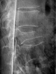
Sunday, April 9, 2017
65 year old with back pain
This image is from 65 year old with complaint of back pain after having week long cold with significant amount of coughing. Pain is general ache, but sharp with rotation and bending activities.
Please make one comment of things you see in this image. Comments can be of things that are normal or abnormal.

Please make one comment of things you see in this image. Comments can be of things that are normal or abnormal.

Thursday, April 21, 2016
Please read this article about postoperative musculoskeletal ultrasound.
Comment on one important "take home" point that you learned about ultrasound after reading this article.
http://e-ultrasonography.org/upload/usg-15006.pdf

Comment on one important "take home" point that you learned about ultrasound after reading this article.
http://e-ultrasonography.org/upload/usg-15006.pdf
Sunday, April 17, 2016
This is a chest radiograph of a 50 year old male who started having pain with deep breathing and coughing following returning from a trip to Italy. Reports pain began after lifting heavy luggage at Eisenhower International Airport upon return home. View radiographs and describe what you feel like you may see and also add one differential diagnosis pathology that you may need to rule out with his history.

Wednesday, April 6, 2016
This should be an easy image to start with. 16 year old female with neck pain following MVA.
On the blog thread, each participant should add one item. Choose from the following. Symptom she would be potentially having, a fact about the injury she has incurred, what is normal or abnormal about the images shown.

On the blog thread, each participant should add one item. Choose from the following. Symptom she would be potentially having, a fact about the injury she has incurred, what is normal or abnormal about the images shown.

Monday, April 20, 2015
Foot Pain
Please see the image below of patient with primary complaint of medial foot pain. Patient was training for marathon and when jumping off of a curb to pavement felt a "pop" while training. they have had foot pain ever since. Each student should add something to the thread. It does not need to be a positive finding that you add it could be something describing a normal finding.

Friday, April 3, 2015
16 Year Old Male with Knee Pain
This is a 16 year old male with medial knee pain, swelling and loss of range of motion. Insidious onset. Started hurting after beginning basketball practice. Can walk FWB but this seems to cause more swelling and discomfort. Rest and decreased WB help. View the AP image and each member of the class note one thing about the image. You are welcome to make a guess as to what you think the problem is. Uses ABCs to make comments regarding this image. You should not need to make the same comments as long as everyone sticks to only one comment about the image.

Monday, April 14, 2014

This 17 year old male football player injured his shoulder following being tackled during football game. Based on your impression of this image what are the possible injuries that could be shown here? What other injuries might you want to rule out based on his mechanism of injury? Based on your impression how would you proceed with therapy during the first several weeks if he came to see you one week after this injury?
Thursday, March 27, 2014
Read the article by Love et al in the journal Radiographics the concept of radionuclear imaging is discussed. Describe one thing that you learned about radionuclear imaging from this article, and how it may relate to a patient you have seen during one of your clinicals!
Wednesday, April 17, 2013

This is a 21 year old male with complaints of shoulder and neck pain. Carefully review your ABC's and see if you notice anything wrong. What type of image is this? Why was it taken? What would be his symptoms? Describe a few and let your other classmates chime in as you progress.
Please complete the post 4/24/2013.
Wednesday, April 3, 2013
Thoracic spine radiograph

From the viewing the image above you can clearly see that it is of the thoracic spine. However look closely and see if you notice anything unusual! Use your ABCs of evaluating radiographic images and tell me as much as you can about this image. Why did they take this image? Is it male or female? What age is the subject? Is the image AP or PA? How can you tell each of these or can you. You do not all need to comment on everything you see, just pick out one or two. If you disagree with your classmates that is ok, just explain why! Each of you should comment about something and please dont just use everyone elses prior comments. Go out on a limb and make a judgement on your own. Its ok if you are not right. Viewing and interpreting images in a skill and art! Even the best radiologists miss the mark sometimes! Good luck!
Tuesday, April 17, 2012

This image is of a male defensive back who injured shoulder after an arm tackle in recent game.
He felt pain and immediate dead arm which lasted 5 min. He has since had pain with ER and abduction movements. Additionally he feels as though his shoulder will give out.
What view are you looking at? What pathology exists and how can you tell?
Make sure you look very closely at the outlines of bony structures!
If he is now a week out - what should you be doing regarding therapy?
How conservative or aggressive should you be?
Wednesday, April 4, 2012

Tuesday, March 20, 2012
Read the article by Love et al in the journal Radiographics the concept of radionuclear imaging is discussed. Describe one thing that you learned about radionuclear imaging from this article, and how it may relate to a patient you have seen during one of your clinicals!
Tuesday, April 26, 2011
Knee Injury

This is going to be a tough one. 20 year old female who injured knee playing basketball when she landed from a rebound with knee in hyperextension. Immediate swelling and giving way following injury. When you view this image where is the abnormality located on the radiograph? Where is the injury located on the image? What is the actual diagnosis? This is classic radiology 101! Hint - you will not find this one in your text! Will require some internet searching!
Good luck!
Good luck!
Sunday, April 17, 2011
Scoliosis
This week you will read an article called "Scoliosis Imaging: What Radiologist Should Know". If you have seen a patient for scoliosis which of the various types of scoliosis did they have? Which radiographic views were, or should have been taken for the given type? If you have yet to see a patient with scoliosis, what did you learn from this article that will help you when you evaluate back patients in the future?
Saturday, April 9, 2011
Posterior Shoulder Pain

This is a adult with complaints of chronic shoulder pain. Pain at activity, rest, and a noticable increase in symptoms at night. Look closely at the shoulder and tell me what you see that may be pathologic and where is it located.
Monday, March 21, 2011
Basic Radiographic Imaging

Which of the following images do you feel better shows the injury that has occured at the ankle? Is this image of an adult or adolescent? How can you tell? How would you describe the position used to take this image? What would be some symptoms this patient would have? How would they present to therapy?
Subscribe to:
Posts (Atom)