Thursday, March 15, 2018
Spine Pain
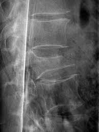
This radiograph is from an elderly woman who accidently fell down a flight of stairs. She has had persistent back pain since that time. Each student should add one piece of information to this case. It can be to describe normal or abnormal things on the radiograph, a finding about whatever you think the diagnosis of this radiograph is, what is a symptom, or what you would do during physical examination. Each blog post should be a different piece of information to lead us toward an accurate diagnosis.
Subscribe to:
Post Comments (Atom)
The first thing I notice is the obvious vertebral body height loss. The vertebral height loss, along with the traumatic incident and patient's age makes me believe this is a compression fracture.
ReplyDelete- Chris Truong
The patient may present with kyphotic posture due to the wedge shape of the fractured vertebrae. Another symptom the patient may present with is increased pain with standing or walking but may decrease with laying down or resting.
ReplyDelete-Senda Vu
I agree with Chris, the first thing In notice with the image is the loss of vertebral height at that segment. Additionally, the asymmetry of the disc space in the segment below. One thing I would look into is how the patient is moving. What motions are restricted, where is the motion coming from, etc.
ReplyDeleteIn my student opinion, I believe I see a pars fracture below the compression fracture. This could lead to a spondylolisthesis.
ReplyDeleteThis comment has been removed by the author.
ReplyDeleteI would examine the convexity of the spine, posture and stance initially. Next, I would look at AROM and gait. The exam would end with light palpation of the vertebrae and of the surrounding paraspinal muscles. She should also be questioned about pain with coughing/sneezing and changes in her overall height.
ReplyDeleteI would look for M/L curves (scoliosis) during my physical exam in addition to what has been mentioned previously. On the two vertebral segments below the crushed one the inferior and superior faces of the vertebral body are visible indicating that they tilted away from mid-line.
ReplyDelete- Adam Veenis
I would start with getting an in depth medical history including information on previous injuries/images in regards to the spine and other diagnoses such as osteopenia/osteoporosis. I think the red flag questions, like Dave mentioned about pain with coughing and sneezing, are really important here. I would also ask questions to determine neurological involvement and part of my exam would include thorough attention to deep tendon reflexes and sensation.
ReplyDelete-Dustyna Roman
What stands out is MOI and pt being elderly female. I agree w/Chris on compression fracture. I had the benefit of seeing a pt on my current rotation who fell and she ended up w/L2 compression fracture. Her fracture was a wedge fracture. As in this image the front of the vertebral body collapses but the back does not forming a wedge.
ReplyDeleteIn addition to the neuro screening that Dustyna mentioned, I would also try to rule out UMN involvement and cord compression by asking about B/B changes, check for Babinski, Hoffman's, Clonus testing, and ask about bilateral symptoms (quadrilateral symptoms not likely with level of injury).
ReplyDeleteMatt Broeckelman
ReplyDeleteI would first educate this patient on back precautions. Teach her to log roll, and to advise her not to lift over 10 lbs, and not to bed forward and twist simultaneously. Also would look into bracing if necessary, to consult with OT about a reacher. In the future, proper hip hinging and squat mechanics should be taught to her to lessen the load on the injured area of the spine. I had a patient similar to this who transitioned from SSB to HH PT care. We recently d/c her with not limitations, she did have her husband as a main caregiver.
First and foremost, I would be thorough on my screening of red flags. Several were mentioned above, but I would also ask about saddle parasthesia. The MOI was traumatic, but I would rule out night pain and unexpected weight loss as well. A previous student mentioned a thorough subjective history, but I think this is an important area to look into further. How long ago was the fall? When were radiographs taken? Are symptoms global or local? How would the patient describe the pain? Does she have any numbness/tingling? It may also be beneficial to have the patient explain the home set-up, particularly the stairs. Are there railings? How many steps? How high are the steps? I would also assess balance in my evaluation. This will not lead me to a diagnosis, but it may be a good indicator on what to work on in therapy, especially considering her MOI was a fall.
ReplyDelete-Kambry Porter
Based on her age and MOI, I'd carefully assess for any other injuries that could have occurred with a fall down the stairs. I've seen a fair share of people with concurrent head injuries (even minor and easily missed symptoms), as well as something simple like a wrist or elbow fracture/dislocation from trying to catch themselves during a fall. Even if all other injuries were ruled out, which I doubt based on her age and MOI, I would throughly examine cognition and follow up with family members to validate any changes they might have seen since her fall. A lot of signs/symptoms of a brain injury, however mild, can take time to appear or evolve and can easily be missed in the acute stages of injury. It's something that needs to be continually monitored throughout treatment as well, even if it's not the center of the primary treatment.
ReplyDeleteSpinal fractures/disc degeneration/any low back injury resulting from trauma are likely to leave the patient in a significant amount of pain during particular bending and twisting movements as explained by others above. One of the more long term complications I would be concerned about is a high risk for decreased physical activity, activities of daily living, hobbies, etc. This will impact the patient's life drastically requiring them to receive more caregiver assistance and decreased general health w/o interventions to prevent such issues. I would work with the patient to come up with ideas for physical activities the patient would be able to perform w/o increasing pain in order for them to stay active and avoid secondary complication d/t decreased activity tolerance. They need to stay as active as possible to maintain their muscle strength and conditioning.
ReplyDeleteMOI and imaging point to compression fracture. D/t pt's age and sex this puts her at risk for osteoporosis, therefore assess for other injuries (if she fell on tailbone, braces with arms, etc). I would perform a through neurological exam and assess for red flags, weakness on LE, sensation changes, spasticity/tone. I would determine PLOF and perform a home screening to assess for home modifications if needed, and educate on spinal precautions.
ReplyDeleteSonia Sanchez
The first thing I noticed is the vertebral height loss in the image that was also noticed by many of my other classmates, I agree with a compression fracture of a potential diagnosis, especially since it is an elderly female. Due to this image the first thing I would do is obtain previous medical information, specifically previous medical history due to age and possibility of osteoporosis contributing to the cause. If osteoporosis is part of the patient's medical history that would also change some of my interventions for this patient due to excessive loading potentially creating more injury to the patient (as Dustyna stated above). I would also assess if the patient would need a lumbar support brace during healing to ensure the patient would not further exacerbate the injury during ADL's due to potential cognitive decline, potential osteoporosis diagnosis due to age and deceased estrogen in elderly women, and/or to help decrease the pain by stabilizing the spine during recovery. Along with the lumbar support brace assessment, I would assess the patients gait and ability to do stairs to see if the patient would need an assistive device for ambulation since the MOI was falling down the stairs. First and foremost I would want the patient to be safe during ambulation whether it be on stairs, curbs, or during gait indoors or uneven surfaces. Obtaining the story about how the MOI happened and assessing her gait would make sure the patient is equipped with the amount of support she would need to be safe.
ReplyDelete-Emily Dreifurst
On the bottom 2 vertebra in the image they seemed to be either turned on an angle like you would see in scoliosis or wedge shaped form side to side, due to the fact the top and bottom are visualized in an elliptical pattern vs is a straight line as demonstrated on above vertebrae. You would need to have additional images including A-P in order to visualize what is going on for sure. You would also want to get the best history you could from this pt and at her age I would bet there would be other images in the past the would help with progression.
ReplyDeleteI would obtain a thorough medical history to see if falls have taken place before and if there was an issue going on previously. As mentioned, I would perform a neurological exam, check for muscle weakness and potential red flags. I would also discuss alternatives to using the stairs if at all possible to avoid another fall.
ReplyDeleteRyan Faflick
I agree with my classmates in that this radiograph demonstrates a lumbar compression fracture. During the examination of this patient, I would do all the neurological screening as mentioned by others above, but would also make sure to assess the sacrum. During her fall, she likely landed directly on her rear end. This could have also caused trauma to the sacrum, in addition to the lumbar compression fracture. Although the SIJ is fused after approximately 60 y.o., the sacrum could still be positioned incorrectly or possibly fractured due to the fall. The sacrum can refer pain to the low back as well, so it should be assessed in this case.
ReplyDelete-Kelsey Darnell
As previously mentioned, this is likely a compression fracture, which could lead to disruption in surrounding tissues like ligaments affecting stability and possibly inhibiting her core. I would like to check how she is engaging her core muscles, primarily the lower abs, and work on strengthening them to be able to progress with decreased risk of injury.
ReplyDeleteIn addition to what my classmates have mentioned above about screenings and assessment, I would be curious as to what caused the fall in the first place. Whether it was dizziness, impaired coordination, muscle weakness, or a simple LOB it would be important to address what lead to the fall as well as the injuries the fall caused.
ReplyDeleteFirst of all, I think it would be important to confirm if the compression fracture is stable or unstable prior to PT treatment to prevent further damage. I also think that taking a close look at balance and gait is huge in this lady, as it has been briefly mentioned by other classmates. Examining the balance and stability of the patient could be helpful in understanding her fall causing this injury and also her risk for encountering falls in the future. Examination of gait helps give us an idea of the areas of weakness and limitations in range of motion, therefore guiding us to a successful treatment plan. The plan should be designed to address any observed gait deviations, weakness in her core and/or BLEs, or balance deficits, ideally resulting in improved gait and stability therefore reducing the risk of falls.
ReplyDeleteMelissa Daughty
I think knowing the patient's body type and how much of an active lifestyle she lives. Knowing this can help guide the treatment in the right direction as well as give us some details on overall health.
ReplyDeleteI would like to know more about what caused her to fall, as treating this underlying issue will be important to help prevent further falls. Also more about the fall itself. Did she fall forward or backward, and how did she land. The answers to these questions will likely lead to other areas that need to be addressed. Since the MOI is a fall down an entire flight of stairs, likely other trauma occured. To my eye, there also appears to be a displaced endplate. This could have occurred as some of the compressive force was converted to an anterior shear force from a rapid flexion motion common with falls. This pt may also not be appropriate for bracing if she had preexisting or increased kyphosis caused by the fx, loss of extension mobility that the brace would promote, or inability to tolerate the confinement. Education will play a key role in this pt's rehab as other people discussed above.
ReplyDeleteI agree that this patient does present a compression fracture, along with a posterior slipped disc and possible DDD. Just by looking at this x-ray, this patient would probably present a lot of swelling, instability, neuropathy, and a kyphotic posture.
ReplyDeleteThe one thing I would definitely pay close attention to would be if the patient presents myotome weakness because that could indicate a possible spinal lesion due to the injury.
If the patient does not present any improvement after a few weeks of therapy, I would definitely recommend her to see a specialist for further imaging.
David Tuong
I agree with the information above and after obtaining a detailed history of falls, health, body type, activity level and any precautions that the MD may have given her, I would want to watch how she gets up/down out of chair and watch her walk. Since it is likely we will be unable to complete all the proper manual muscle tests due to pain or restrictions from the MD, seeing the pt ambulated in the clinic can give you an idea of what muscles we will need to focus on. My first goal would be to get pain under control and increase ROM but we need to address underlying issues including weakness of hip/ core stabilizers to ensure safe return to ADL’s/recreational activities. The evaluation, I may not do a lot of actual therapeutic exercises, I would educate ALOT, and try to help reduce pain. After appointment I would call the MD to clarify anything and touch base with anything extra they wanted to add that the patient may have forgotten.
ReplyDelete~Courtney Simon
In this radiograph, I noticed the decreased lumbar lordosis, the decrease and abnormal shape of the vertebral body height and the decreased disc space between the vertebral bodies superior to the fracture site. I found a research article (https://www.ncbi.nlm.nih.gov/pmc/articles/PMC4056125/) regarding imaging of vertebral fractures and found a diagram to represent Genant’s method of grading vertebral fractures by the extent of vertebral body height reduction. Looking at this radiograph and the diagram, I would say this particular patient has suffered from a moderate-severe fracture.
ReplyDeleteGiven the woman’s age and the mechanism of injury, I believe this is a radiographic image showing a vertebral compression fracture. To start my evaluation of this patient, I would first do a thorough medical history better understand the patient and how this incident occurred (could it be osteoporosis, trauma from the fall, or possibly a pathologic fracture?) and contact the doctor to clarify. I would ask her pain level and if she could describe the pain that she is feeling. With vertebral compression fractures I would expect to hear about pain in the mid-lower back and possibly numbness and tingling or even losing control of bowel/bladder. If numbness and tingling are present, this could mean compression of the nerves at the fracture site and loss of bowel/bladder could mean that the fracture is pushing on the spinal cord itself (red flag – send patient to ER). I would then, so long as symptoms allow, do a visual examination of her posture (increased kyphosis? Forward head?). Then I would begin to perform neurological tests and assess ROM/strength as well as transfers and gait.
This is an unstable compression fracture. The superior-anterior corner of the vertebral body is displaced from the rest of the vertebral body. There appears to be a linear zone of compression inferior to the superior vertebral endplate, which indicates an recent (<2 months old) compression fracture.
ReplyDeleteI would first screen for any red flags, obtain a full patient history and any symptoms the patient may be having such as pain radiating into legs, loss of sensation, localized pain and specific ROM active or passive. Based on the image the shape of the vertebrae appears to have slipped and a compression fracture is likely. I would be careful not to include any extra load to site due to suspicion of fracture but when pain free would incorporate, abdominal strengthening, stretching and proper mechanics with functional activity. Prior to this point I would focus on pt education and potentially bracing if necessary.
ReplyDeleteMichael Edwards
The first thing I noticed was the decreased height of the vertebral body in the center of the radiograph, indicating a fracture and/or degeneration. The vertebral body below seems to be degenerative on the inferior side. Along with checking for read flags and gathering objective info, I would ask about employment, ADL's and hobbies to assess PLOF and patient interests. I could use this info to write specific goals and provide motivation for the patient to return to PLOF. It would also be important to teach her how to move safely during work/ADL's/hobbies, especially if this is what caused the fall.
ReplyDeleteLani Martin
How old is this image? How old is this injury? Does taking big breaths hurt? What position is she able to sleep in? What movements are painful? What movement impairments are identified? What are her contributing factors to her movement impairments? Side-bending hurt worse than rotating? Etc... By just the alignments of the anterior vertebral body line, posterior vertebral line, and spinolaminar line I would expect some neurological signs and symptoms. The segment with obvious deformity also appears to be in an extended position compared with the inferior segment.
ReplyDeleteI agree with the majority of what my classmates have mentioned above: compression fx; in depth subjective history to pick out any red flags; standing assessment of movement; scoliosis screen; definitely neurological assessment; etc. After that is complete I would look at her function (bed mobility; ambulation; any assistance being required at home or wherever this patient may live). I would assess how this patient is negotiating stairs to assess safety in hopes to prevent future falls with this activity. I would also perform balance testing for recommendations on an AD if one is not being used and needs to be.
ReplyDeleteI agree with most of my classmates that the imaging represents a compression fracture with possible degenerative changes in the lumbar spine. First, I want to know how long ago the injury occurred and how recent the imaging is. Screening for red flags and thorough subjective history about the patient and this trauma is very important and would also be one of the first things I do to help determine if there is a need for referral or if the patient is appropriate for PT. I agree that based on this patient’s age and fall it would be important for thorough screening of cognition especially over time because a head injury could evolve and worsen over time. I would use the subjective history and MOI to determine possible factors leading to this trauma and identifying what PT should address to help prevent further injury and future falls. If this patient is not at PLOF they may benefit from other disciplines to help return to PLOF if appropriate.
ReplyDeleteKassidy Simmons
I agree with my fellow peers and their recommendations for this patient (I'm reminded of how smart everyone is.) In addition to what my classmates have said, I want to ask this lady about her nutrition. Considering her age and high risk for osteoporosis/osteopenia I want to make sure she is eating foods high in protein and Vitamin D, like dairy, fatty fish, and a good amount of dark leafy greens for good measure. She should be educated on what to limit/stay away from, like caffeine and alcohol which can interfere with calcium absorption and lead to bone loss. I also would be curious to see which exact medications she is on to make sure there are no contraindications and she is getting the supplements she needs. If she required further help/instruction in this area I would refer her to a dietician/pharmacist as needed.
ReplyDelete-Alice Hartman
If this was a recent fall and I was seeing this patient in the inpatient setting: I will first do a mini mental test (ask her name, if she knows where she is at, the day/month/year). I would ask her if she lives by herself, if she drives or if there is someone that will be helping her with that, how many stairs/steps are in her house and if they have handrails, was she using an assistive device prior to this, I will also ask her to rate her pain. I will also walk with her to see if she is able to walk an appropriate distance for her to access her bathroom/kitchen/bedroom at home. Lastly, the only different thing I would do during the examination that my classmates haven't mentioned, is that I will do MMT of her upper extremities as well (maybe she fell from the stairs due to upper extremity weakness/shoulder pain/etc).
ReplyDeleteElvia Barraza
Loss of overall height up to 6 inches often come with compression fractures. I would subjectively assess her height pre-injiry and take height measurements after to determine the extent of the fracture. Mikalea Bunyan
ReplyDeleteAs has been previous stated by my classmates, this appears to be a compression fracture due to the shear loss of vertebral height. I had a patient with this same diagnosis in the thoracic spine a few weeks ago and it was very painful for her. She had limited rotation, significant muscle spasm in bilateral paraspinals and very limited flexion and extension range. One thing she really benefited from was manual lumbar/pelvic traction with a gait belt around her upper legs near the groin area and then her legs supported on a swiss ball. Following about 10 minutes of this treatment she frequently had symptomatic relief. We focused heavily on introducing rotation exercises such as lower trunk rotations on a swiss ball and seated twisting within a pain free range.
ReplyDeleteAs everyone has stated, I noticed there is a loss of vertebral height. I would ask about pain descriptors to help lead to me making a possible PT diagnosis with pain describers including stabbing and non-radiating. During the examination, I would assess overall standing posture since this patient is an elder. I would examine if this patient has a scoliotic curve as this can also lead to a compression fracture. In a chart review, I would look for bone density information as osteoporosis could also contribute to the risk of a compression fracture.
ReplyDelete-Kailey Thach
I would agree with my classmates that this image appears to show a compression fracture which is a common result considering the MOI and that the patient is an elderly women. I would want to check for pre-existing conditions such as osteoporosis and assess for any red flags or neurological symptoms. I would also want to assess this patient's breathing and educate her to make sure she is breathing properly in order to prevent pneumonia. The elderly patient population is more at risk of pneumonia so it would be important to educate her to be aware as increased pain with breathing could put this patient more at risk. She may also need some education on ways to reduce pain when coughing or sneezing.
ReplyDelete-Kendra Hallacy
As most of my classmates have stated, I agree that the image shows a loss of vertebral height which could be a contributor to the apparent compression fracture. I think it would be important to know when the fall occurred and whether it was acute. I would like to know the date the imaging was performed to confirm they were up to date. If the fall was acute, I would make sure to perform a cognitive screening to rule out any deficits that may not be apparent with normal musculoskeletal evaluation.
ReplyDeleteThen, I think it would be important to determine if this fracture were stable or unstable prior to performing interventions to prevent the possibility of causing further damage in the case of an unstable fracture.
I would like to know what caused the fall, or if she has had prior falls, and how many. I want to assess her balance and stability with functional tasks, as well as examine her gait to give me a solid baseline of her deficits with functional activity. One more important area of my evaluation and treatment would be proper and safe stair negotiation so that she can ascend/descend stairs to enter/exit home using prevention strategies learned to prevent another fall from occurring.